

Maggiori informazioni sul libro
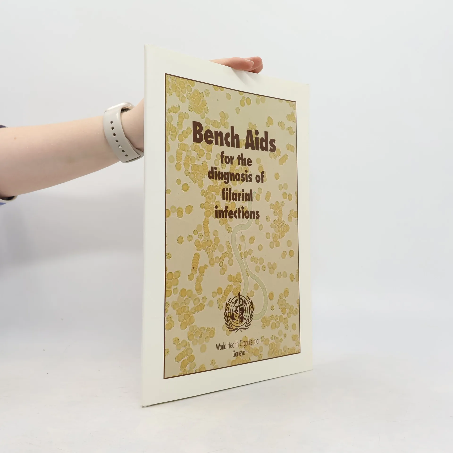
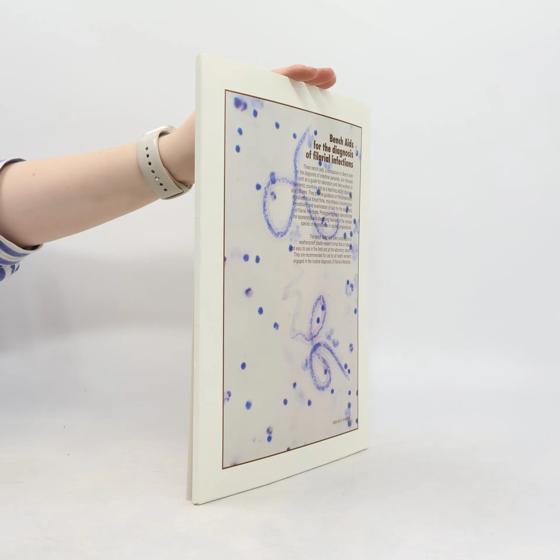
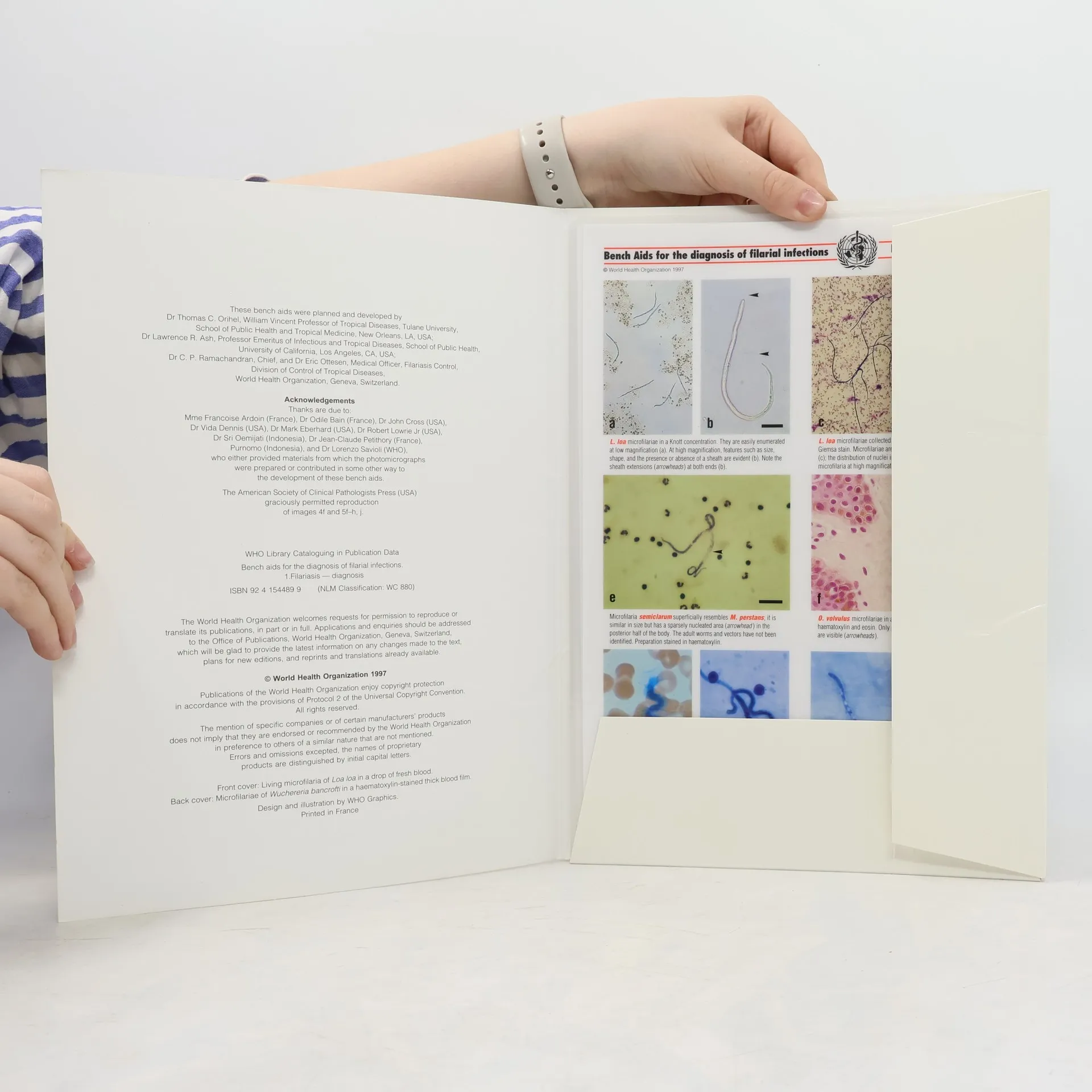
This practical tool aids in diagnosing filarial infections through 46 full-color photomicrographs that showcase the distinctive features of various microfilariae species in different stained preparations and magnifications. Each image includes a measuring bar and a brief explanatory legend highlighting key characteristics for accurate diagnosis. The species featured are Wuchereria bancrofti, Loa loa, Brugia malayi, Brugia timori, Mansonella perstans, Mansonella ozzardi, Onchocerca volvulus, and Mansonella streptocerca, along with images of common artifacts that may complicate diagnosis. The six plates are designed in a durable plasticized format, serving as a guide for laboratory and field workers in endemic regions, as well as a teaching resource for students and trainees. The objective is to enable microscopists to achieve quick and precise diagnoses while minimizing errors. To support this aim, the bench aids include essential laboratory instructions alongside high-quality images, detailing the preparation of thick blood films, Giemsa and haematoxylin staining, membrane filtration, the Knott concentration method, and tissue examination techniques for detecting microfilariae in the skin. Additionally, a comparative summary outlines the geographical distribution, vectors, habitat, periodicity, and key diagnostic features of the most common human filarial parasites.
Acquisto del libro
Bench Aids for the Diagnosis of Filarial Infections, World Health Organization, Lawrence R. Ash, E. Ottesen, Thomas C. Orihel, C. P. Ramachandran
- Lingua

- Pubblicato
- 1997
- Rilegatura
- (In brossura)
Metodi di pagamento
Ancora nessuna valutazione.
- Titolo
- Bench Aids for the Diagnosis of Filarial Infections
- Lingua
- Inglese
- Editore
- World Health Organization
- Pubblicato
- 1997
- Formato
- In brossura
- Pagine
- 6
- ISBN10
- 9241544899
- ISBN13
- 9789241544894
- Serie
- Categoria
- Tag
- Salute & Medicina, Manuali e guide, Scienza, Medicina, Tecniche di laboratorio, Medicina Tropicale
- Descrizione
- This practical tool aids in diagnosing filarial infections through 46 full-color photomicrographs that showcase the distinctive features of various microfilariae species in different stained preparations and magnifications. Each image includes a measuring bar and a brief explanatory legend highlighting key characteristics for accurate diagnosis. The species featured are Wuchereria bancrofti, Loa loa, Brugia malayi, Brugia timori, Mansonella perstans, Mansonella ozzardi, Onchocerca volvulus, and Mansonella streptocerca, along with images of common artifacts that may complicate diagnosis. The six plates are designed in a durable plasticized format, serving as a guide for laboratory and field workers in endemic regions, as well as a teaching resource for students and trainees. The objective is to enable microscopists to achieve quick and precise diagnoses while minimizing errors. To support this aim, the bench aids include essential laboratory instructions alongside high-quality images, detailing the preparation of thick blood films, Giemsa and haematoxylin staining, membrane filtration, the Knott concentration method, and tissue examination techniques for detecting microfilariae in the skin. Additionally, a comparative summary outlines the geographical distribution, vectors, habitat, periodicity, and key diagnostic features of the most common human filarial parasites.