Gerhard Wanner Libri


A concise and authoritative introduction to scanning electron microscopy in the biological sciences In A Practical Guide to Scanning Electron Microscopy distinguished electron microscopist Gerhard Wanner delivers a practical handbook for biological scientists working with microbial, plant, and animal cells and tissues, enabling them to successfully apply scanning electron microscopy (SEM) to their object of study. The book begins with an introduction to the principles of electron microscopy and the operation of electron microscopes before moving on to describe the preparation and mounting of specimens. It also explores the process of recoding images and their subsequent analysis, along with a wide range of advanced microscopy techniques, including cryo-SEM, FIB-SEM tomography, and stereo-SEM. Scanning Electron Microscopy in the Biosciences contains hundreds of carefully selected microscopic images, as well as hands-on, step-by-step guidance required to perform a successful TEM experiment. Readers will also Perfect for cell biologists and microbiologists, A Practical Guide to Scanning Electron Microscopy in the Biosciences also belongs in the libraries of neurobiologists and biophysicists.
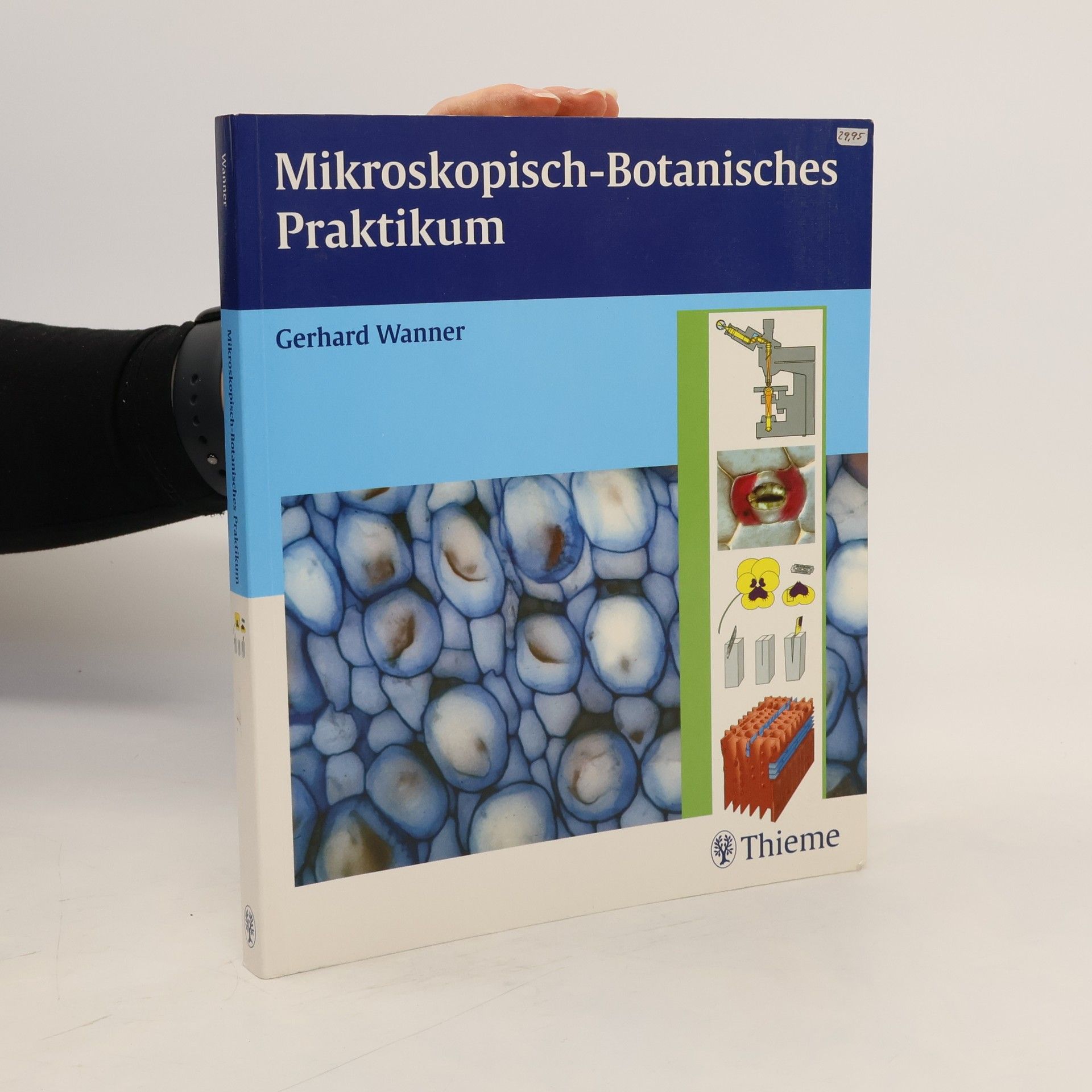
Präparieren, Erkennen, Zeichnen - Dein Leitfaden durchs Praktikum!Hier findest Du alle üblicherweise im Kurs behandelten Themen der mikroskopischen Pflanzenanatomie:- Der richtige Umgang mit dem Lichtmikroskop- Grundlagen der Elektronenmikroskopie- Wichtige Schnitt- und Färbetechniken- Interpretation von Gewebeschnitten- Zeichnen von Zellen und GewebenInhaltlich abgeschlossene Doppelseiten:Ideal für den Kurs und zum Mikroskopieren- Brillante Fotos illustrieren die pflanzlichen Strukturen und wecken Interesse für das Fach- Praktikumsrelevante Inhalte: Kaum #Ballast# durch Grundlagenwissen zur Botanik
