Covers the entire musculoskeletal system with an array of magnetic resonance (MR) images as well as four-colour gross anatomic dissections, four-colour arthroscopy and four-colour, three-dimensional imaging techniques and final renderings.
David W. Stoller Libri
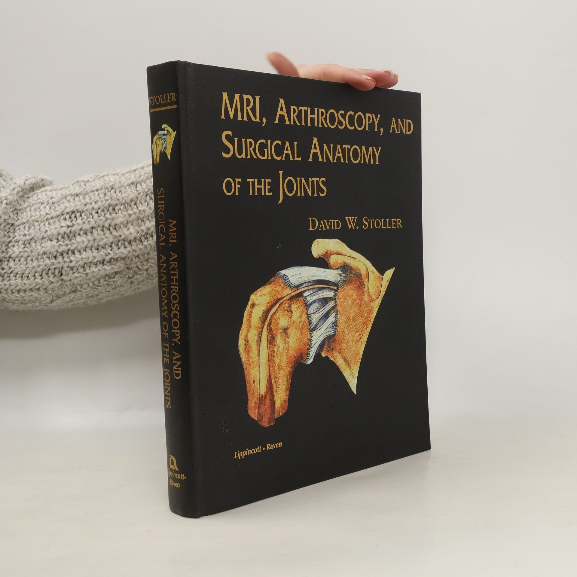


This atlas vividly depicts the anatomy of the shoulder, ankle, hip, knee, wrist, and elbow as seen in magnetic resonance imaging, arthroscopy, and surgical dissection. Featuring over 500 MRI scans and over 200 full-color arthroscopic and surgical dissection photographs, the atlas shows direct correlations among MRI, arthroscopy, and surgical anatomy. MRI scans of cadaver joints are presented alongside arthroscopic and surgical dissection photographs of the same cadaver specimens. Each chapter begins with a commentary by an eminent orthopaedic surgeon on arthroscopy and surgical anatomy. MRI, Arthroscopy, and Surgical Anatomy of the Joints is also available on a multimedia CD-ROM. See Media section for details.
Stoller's Orthopaedics and Sports Medicine: The Knee
Includes Stoller Lecture Videos and Stoller Notes
- 1000pagine
- 35 ore di lettura
This comprehensive multimedia package offers an in-depth exploration of knee anatomy and treatment from a leading expert in orthopaedics and sports medicine. It includes a print book, engaging lecture videos, and supplementary "Stoller Notes," providing a rich learning experience for students and professionals alike. The combination of visual and written resources enhances understanding of complex concepts related to knee health and injury management.
Wer radiologisches Praxiswissen in kurzer, prägnanter und übersichtlicher Form sucht, dem hilft der PocketRadiologist in der Kitteltasche. Denn hinter dieser Buchreihe steht eine renommierte, internationale Herausgeberschaft und ein Konzept, das in der Radiologie einzigartig ist: In jedem Band der Reihe finden Sie die 100 wichtigsten Diagnosen des jeweiligen radiologischen Teilgebiets. Jede der 100 Diagnosen ist nach dem gleichen Schema dargestellt: Detaillierte Aufnahmen und Zeichnungen Die radiologisch wichtigsten Fakten zum Krankheitsbild Befunde der Bildgebung - CT, MRT, konventionell… und Empfehlungen für die rationelle Diagnostik Differenzialdiagnostik Ätiologie und Pathogenese Klinik und Verlauf Ausgewählte Literatur Diese stringente Darstellung macht es dem Radiologen leicht, seine Verdachtsdiagnose akku-rat und schnell zu bestätigen oder auszuschließen. Spezialistenrat aus der Kitteltasche!